A biopsy of the mass was performed and diagnosed histopathologically as a follicular cyst. The condition is caused when the puppies are developing as embryos and the spinal column that develops from a neural tube does not part from the skin resulting in a bulge or cyst along the back.

Hello My Dog Is A 6 Year Old Pomerianian Shih Tzu I Recently Noticed Three Small Bumps On Him One On His Side On On His Tail And Petcoach
These masses may be found anywhere on the body.

Cutaneous cyst shih tzu. Most Common Shih Tzu Eye Problems. Sebaceous glands are microscopic structures found just beneath the skin surface. If the cyst is infected it may be red inflamed and painful.
A sebaceous cyst may be firm or it may feel like it is filled with fluid. A sebaceous cyst typically appears as a small raised well-defined round structure in the skin. False cysts may be formed by hemorrhage or trauma that leads to tissue death.
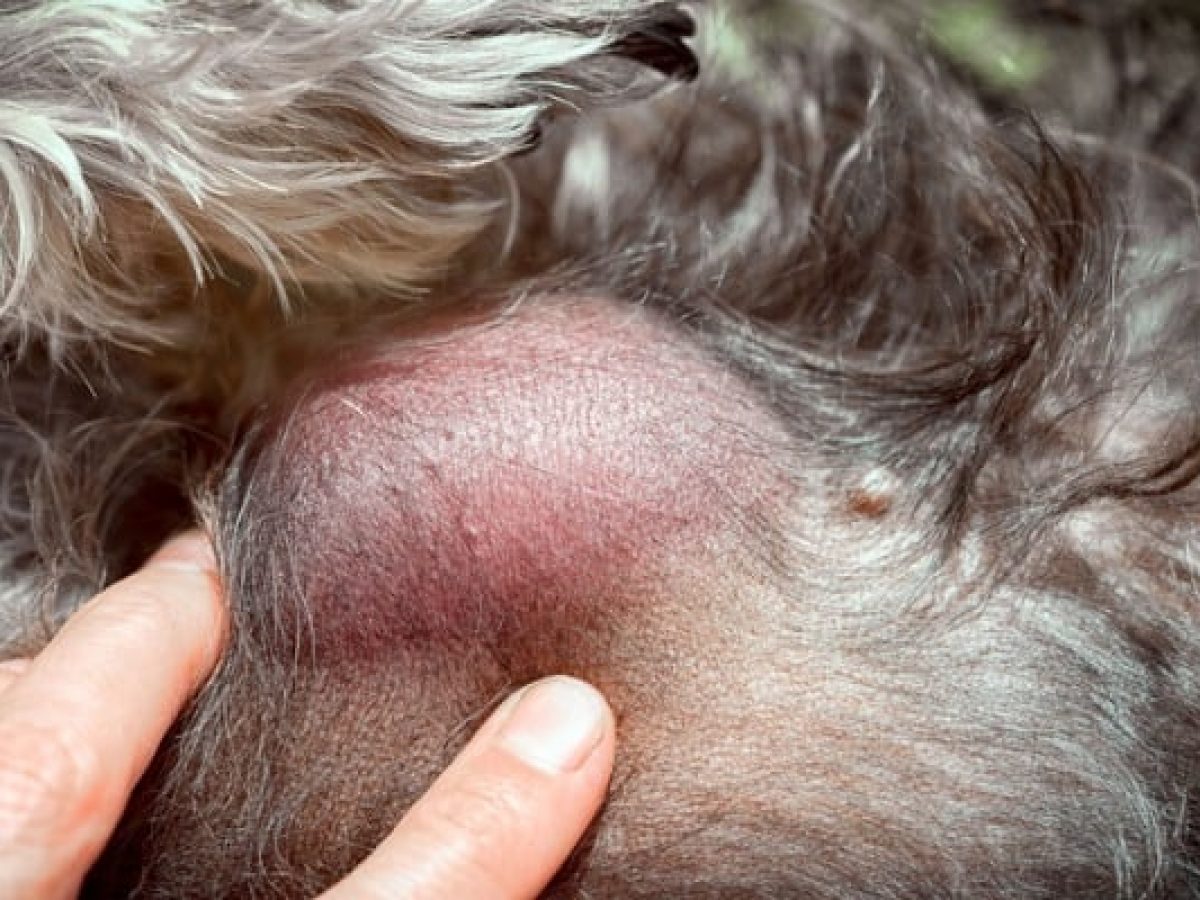
I thought Id check with the community to see if maybe somebody had any success treating them. A dorsal tail mass 1 x 12 cm was observed on a 10-year-old castrated male Shih-tzu dog. Sebaceous cysts on Shih Tzu skin are usually due to blocked hair follicles causing sebum your dogs lubricating skin oil to collect block and swell the follicle.
There was no significant breed predisposition P 086. From the sinus and the sinus was neither painful or inflamed. No sex predilection has been noted.
Sebaceous cysts are common types of skin cysts that contain sebum a thick oily material normally found in the skin around the hair follicles. Journal of Animal Health and Production 9 147-51. 4950 Apocrine tumors are most common in middle-aged to older dogs.
Given the opportunity to examine an older dog Ill very likely find at least one or two cutaneous within the skin or subcutaneous just beneath the skin surface lumps and bumps. Usually these cysts are solitary but some dogs may be prone to getting several cysts in the same area of the body. Breeds between 8-11 years have a higher incidence.
Can also cause the blockage. Dermoid cysts are rare. Dermoid cysts are complex congenital cysts that form long before birth.
SD 41 with four dogs 35 years old. Damage to the hair follicle from pressure points from a harness lying down etc. The 4 masses were aligned breeds and are usually diagnosed in young animals3 Dermoid.
Sebaceous cysts are common in dogs but unusual in cats with the exception of stud tail on the upper side of the tail. Trim the hair around the opening be careful with scissors we dont want that hair matting on top not allowing it to drain. If it is open the cyst or bulge is still attached to the spinal column however if it is what is known as a blind sac there is no actual connection just the physical cyst.
All the dogs presented with one or more firm or soft cutaneous nodules. Cutaneous apocrine gland carcinomas are uncommon. The age of onset ranged from 3 to 15 years mean 74.
Shih Tzu dogs have very large slightly protruding eyes. Its part of what makes them so cute but its also a health hazard. Can also cause the blockage.
If left alone it might go away without treatment but may recur. In dogs the cutaneous dermoid cysts are most often seen in the Rhodesian Ridgeback Siberian Husky Shih Tzu Boxer and Kerry Blue terrier breeds and are usually diagnosed in young animals. My vet basically said my lil turbo had to live with them.
For carcinomas they include the Old English Sheepdog shih tzu German shepherd. Cutaneous dermoid cysts are most often seen in the Rhode-Deep palpation identified 4 separate masses that varied insian Ridgeback Shih Tzu Boxer and Kerry Blue terrier size from 05 to 1 cm in diameter. The discharge they produce makes for a very accommodating environment for bacteria to breed.
These are far and away the most common benign skin tumors in dogs. They are unlike the cysts he has had on his back. The lobules are filled with a clear fluid.
The lesions can be ulcers nodules lumps plaques reddish patches or areas of scaling and hair loss. Has anybody had any luck treating sebaceous cysts on their Shih-tzu. The Shih Tzu I asked about last week now presents with jelly like cysts which rupture when any pressure is applied.
False cysts are fluid-filled structures that do not contain a secretory lining. Lhasa apso Old English sheepdog Collie Shih Tzu and Irish setters are highly predisposed. Damage to the hair follicle from pressure points from a harness lying down etc.
In most cases dermoid cysts in dogs are associated with multiple vertebral and spinal malformations and hind limb neurologic deficits Jubb et al. If it bursts a white paste-like or cottage-cheese-like material is seen. In dogs cutaneous dermoid cysts are common in some breeds and are usually diagnosed in young animals Burrow 2004Cornegliani Jommi.
Basically a sebaceous cyst is a very large pimple that is harmless to your pet and may feel like a raised bump. The large exposed eyes are prone to many irritations and eye infections. Our Shih Tzu gets skin bumps and rashes due to food allergies.
Weve tried all sorts of lotions and potions and the things that have worked best are grain and poultry-free food hydrocortisone spray for when there is an attack of the itchies and a water pistol to discourage for foot licking My Shih Tzu has terrible skin problems. Some lesions are multilobulated and cystic. Understand the causes.
Sebaceous cysts on Shih Tzu skin are usually due to blocked hair follicles causing sebum your dogs lubricating skin oil to collect block and swell the follicle. In this regard I liken them to the brown spots that appear on our skin as we get older. Most dogs will develop at least a couple of them by the time they are senior citizens.
Use warm compresses and digital massage to draw out the fluid then ivory soap and water or dog oatmeal shampoo to the area twice daily with ten minute contact time before rinsing and tap dry. Predisposed breeds for adenomas include Lhasa apso Old English sheepdog collie shih tzu great Pyrenees chow chow malamute and Irish setter. Sebaceous cysts are benign but can also be mistaken for a malignant tumor called a sebaceous gland adenocarcinoma or a benign mass called a sebaceous.
Malignant Cutaneous Melanoma in a Shih Tzu Mix. Heres how to clean Shih Tzu eyes signs of Shih Tzu eye problems and how to remove Shih Tzu tear stains Canine cutaneous lymphoma can present in quite a variety of lesions. Such growths are common by-products of the aging process.
Jelly like cystsunlike the cysts he has had on his back. The cysts also have fine interlobular separations of connective tissue. The study population comprised six male neu- tered two female spayed and two female intact dogs.
A necrotic lesion 25 x 3 cm developed at the biopsy site 1 week after sampling and failed to respond to 2 weeks of normal saline cleansing and systemic antibiotic administration.
Keratitis symptoms include intense lacrimation photophobia and protrusion of the third eyelid. Benign neoplasms are more common than malignant neoplasms and epithelial neoplasms are more frequent than mesenchymal neoplasms.
Canine Eyelid Masses Acvo Public
A growth on the eyelid should be evaluated by a veterinary professional without delay.
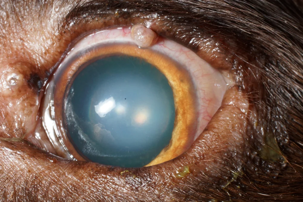
Cyst on eyelid of a dog. Pet eyelid tumor treatment. Repeated pawing or rubbing the eye. Sebaceous cysts are harmless cysts which can appear on the eyelid or any other part of the skin.
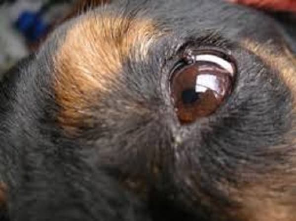
Rougheningthickening of the conjunctiva. I felt it and its hard. Meibomian gland cysts in dogs typically occur in older dogs although they can occur in all ages and all breeds.
A sty is often the name given to a bump on a dogs eyelids that appears similar to an isolated pimple. How do you treat a cyst on a dog. The vet may wish to send the lump away to the lab for analysis to double check the tumor is.
Signs of pain were not evident during. Other signs that your pet may have a tumor of the eyelid conjunctiva or periocular tissue include. Common symptoms of clogged glands are inflammation swelling and pain to the touch.
My dog has a little stye or cyst in his upper eyelid. Pain or vision inhibition can. Multiple pigmented nodules were seen around the skin of the upper and lower left eyelids.
With time sebaceous cysts often rupture and their creamy contents are released. The nodules were brownish to black round soft and fluid-filled. Meibomian glands are sebaceous glands that provide an oily secretion to stabilize the tear film over the cornea Common in older dogs meibomian gland tumors are usually benign but a small.
Both dog and cat eyes may be affected by these but they are more common in dogs. It doesnt look like it could be scratching the eye in any way. While there are many treatments for these cysts many of them will go away on their.
There are different types of keratitis in dogs such as ulcerative infectious interstitial vascular and pigmentary. The lesions if left alone however have the potential to be locally aggressive and disfiguring leading to ocular surface irritation or worse corneal ulceration or infection. Redness swelling pawing at the area crusting of the eyelids and fur loss around the eyes could be signs of trouble.
If you notice a growth on your dogs eyelid it could be whats known as a meibomian gland cyst or chalazion. Many eyelid tumors in dogs are overgrowth of the meibomian gland. It looks like a pimple or bump on the eyelid margin.
Fatty tumors can develop in any breed of dog but larger breeds and those that are overweight or obese are more prone to having them. They typically develop in older dogs but can occur in pets of any age. 7 Common eyelid neoplasms are described in Table 2.
Sebaceous cysts appear as growths on the skin of a dog. Its attached to his lid but sticks out from the lid so it isnt raising the eye lid upward. What can I do for my.
If its a subaceous cyst the bump on your dogs eye may be filled with fluid or solid. When the dog blinks a large eyelid tumor rubs backward and forward over the surface of the eye which can cause inflammation or even ulceration of the surface. However mast cell tumors histiocytomas and.
Apply a warm compress to the affected eye for 10 to 15 minutes 2 to 4 times a day for several days. Eyelid masses can be detrimental to your pets health and quality of life but fortunately most eyelid masses behave in a benign nature and do not result in spread of disease to distant areas of the body. Eyelid growths can be common in dogs.
Health Navigator says these are usually caused by blocked oil glands and they are not due to infection. Again this is down a blockage but this time in a follicle around the eyelashes. Eyelid masses can be detrimental to your pets health and quality of life but fortunately most eyelid masses behave in a benign nature and do not result in spread of disease to distant areas of the body.
Eyelid growths can be common in dogs. Protrusion of the third eyelid. How are these types of tumors diagnosed.
All types must be treated accordingly to avoid blindness. The most common treatment for cysts is surgical removal. Eyelid tumors can be treated a variety of ways depending on the tumor type size and extent.
After applying the compress use your finger to press on the. A sty on a dogs eyelid is caused by an infection in the eyelid gland. These pimple-like growths on your dogs skin are blocked sebaceous glands.
Keratitis in dogs consists of inflammation of the cornea which becomes cloudy and loses transparency. These tumors are tiny slow-growing tumors that form in the meibomian glands of the eyelids. Avoid popping the sty because it is.
In young dogs less than three years old they tend to be caused by a virus papilloma virus and often resolve without treatment. A cyst on the eyelid called a meibomian gland adenoma can interfere with the ability to blink or cause excessive glare. They typically develop in older dogs but can occur in pets of any age.
Fortunately for most dogs the vast majority of eyelid margin tumors are benign so there is little risk for metastases and surgery is usually curative. Are eyelid tumors in dogs dangerous. These tumors are tiny slow-growing tumors that.
Formally known as a hordeolum a sty is ultimately an abscess which either will form a head and eventually burst or will eventually reabsorb. A small superficial or benign tumor can possibly be debulked and treated with cryotherapy using local anesthetic and sedation while a malignant full thickness or large tumor may require general anesthesia and removal. Keeping this in view how do you get rid of a stye on a dogs eye.
Pet eyelid tumor treatment A small superficial or benign tumor can possibly be debulked and treated with cryotherapy using local anesthetic and sedation while a malignant full thickness or large tumor may require general anesthesia and removal of a portion of the eyelid. In older dogs they tend to develop with no obvious cause and grow slowly over. Warts - warts on the eyelid are hairless bumpy and usually the same colour as the eyelid for example pink from a pink eyelid and black from a black eyelid.
7 Most eyelid neoplasms occur in dogs older than 10 years of age with the superior lid affected more often than the inferior lid. Pay particular attention to your dogs eyes for any changes however subtle. Clogged Glands Stye Similar to stye in humans your dog might have clogged oil glands or infected follicles that might look like bumps on your dogs eyelid.
A mass on the eyelid of your dog can be benign or malignant and can occur due to gland issues or a genetic breed predisposition. Styes are very common in humans but in dogs they are actually rare. A sty on a dogs eyelid is caused by an infection in the eyelid gland.
Sebaceous cysts form when the oil gland or duct the opening through which the oil travels through gets blocked and becomes clogged. There are two main types of prostatic cysts in dogs.
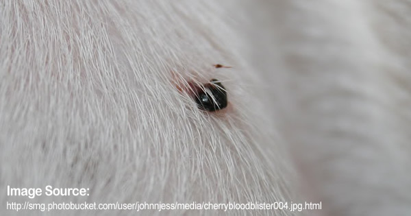
Pet Care Symptoms Blood Filled Lumps Petpremium
With time sebaceous cysts often rupture and their creamy contents are released.
Blood cyst dog. Youll probably find this type of cyst on your dogs head trunk or neck where. Interdigital follicular cysts in dogs are quite common. Carl pops a baseball size sebaceous cyst on a k9 patient.
Sebaceous cysts in dogs are typically smaller than an abscess. They usually appear as blood- or pus-filled red nodules between the toes usually on the front feet and form as a result of excessive friction or trauma to the webbing between the toes. Most sebaceous cysts dont cause problems for dogs so there is not typically a need to remove them unless theyre infected.
The dog may also scratch the cyst or bite the crusts leading to bleeding. If you touch them theyre unlikely to cause your dog any discomfort and should not feel hot or warm. A sebaceous cyst typically appears as a small raised well-defined round structure in the skin.
There are many different types of cysts that. Cysts tend to occur in middle-aged or older dogs and are most commonly linked to breeds such as German Shepherds Boxers and Cocker Spaniels. The tumors invade and erode blood vessels which can cause a considerable amount of blood to seep into the abdominal or less commonly the thoracic chest cavity.
Pet owners will first notice a sebaceous cyst when they discover a raised bump on their dogs skin. It is found in different shapes. Histiocytoma tumors are usually seen on the ears head and legs.
When a dog finally begins to show symptoms they usually present as the following. The sebum is necessary to lubricate the hair. You will either find this extremely gross or extremely awesome if youre the latter- enjoy.
If your dog scratches at the bump it may start to bleed. This time though the open wound had become infected and needed a cleanup and antibiotics. Since dogs with urolithiasis often show signs similar to those with prostatic disease the radiograph should be carefully evaluated for calculi.
Blood-filled lumps are usually caused by physical trauma. Its important to discuss any new lumps with your. Neither type are cancerous however the cysts do need to be removed to ensure the dogs normal bodily functions are able to perform without interference from the pressure of the.
Blood blisters on dogs are usually the result of an injury or trauma of some kind although they can also occur due to a blood clotting problem. In the case of an aural hematoma a dog or cat may be itchy as a result of ear mites or an ear infection. Its not advisable to pop them because it can increase the risk of secondary bacterial infection.
The cyst can then either be allowed to drain by itself into a pan or suction can be applied to remove the contents. It occurs when blood leaks out of a blood vessel. Dermoid cysts are complex congenital cysts that form long before birth.
After the cyst is drained most vets will opt to remove it entirely especially if it has become infected. Follicular cysts appear on or beneath your pets skin. When they are caused by a clogged gland they can be transparent or bluish but some can be dark too.
Sebaceous cysts are common in dogs but unusual in cats with the exception of stud tail on the upper side of the tail. In most cases sebaceous cysts have a size ranging from 5 mm to 5 cm. Intense scratching and head shaking can inflict trauma to small blood vessels in the outer ear.
My dog had a blood filled cyst filled with blood that burst. What is important to note with regard to sebaceous cysts in dogs is that. False cysts are fluid-filled structures that do not contain a secretory lining.
Material strongly positive for factor VIII-relatedantigen is present in the cytoplasm ofround endothelial cells. Pale mucous membranes mouth and eyes Rapid or labored breathing. Your dog has sebaceous glands which are glands in the skin that open into a hair follicle and secrets sebum.
Typically found in dogs with pendulous ears and as the result of an ear infection. Dermoid cysts are rare. Interdigital Follicular Cyst on Dogs.
Cysts abscesses size echogenic texture and changes suggestive of BPH or neoplasia can all be. Depending on the size and type of the cyst the contents that spill out together with blood will vary in color quantity and smell. I flushed it with.
Rapture of the cyst usually happens due to trauma or injury directed towards the cyst. Technically the term cyst is a bit of a misnomer because it relates to a specific type of swelling with a specific type of cell lining and interdigital cysts do not typically have that structure. False cysts may be formed by hemorrhage or trauma that leads to tissue death.
Paraprostatic around the gland and those formed on the gland. My small dog had a bug cyst bigger then a quarter sizer on. The Types of Canine Cysts.
It looks like a strawberry. Technically speaking a cyst is a sac in the skin lined with secretory cells. These cysts are round and have a bluish tint.
Abdominal ultrasound has become indispensable for evaluation of the abnormal canine prostate gland. The incision is then sutured closed and the dog allowed to awaken. Think of a sebaceous cyst like a large pimple sometimes they can grow into visible prominent bumps on your dogs skin.
It popped and is ozzing blood but the hole looks rather large. A tumor found in young dogs that results in a raised red nodule. My small dog had a bug cyst bigger then a quarter sizer on her back.
The ruptured cyst is in most instances less painful. A canine interdigital cyst is a bump or swelling that occurs between a dogs toes. Factor VIII-relatedantigen is detected in blood cysts from a 5-month-oldFig.
Sebaceous cysts can occur anywhere but may be more common along the head neck and trunk. Your veterinarian can examine the. 3a dog and from a 24-month-oldFig.
Possibility of blood in the urine.