Canine heart model and lungs infested with heartworms dirofilaria immitis. The digestive system absorbs and digests food and eliminates solid wastes from the body.

Anatomy Model Heart And Lung Canine
Dog Tail Anatomy The tail of a dog serves many functions such as non verbal communication and as a rudder in water.
Canine heart anatomy. Heart and Major Vessels. Canine Heart - Normal Rostral View. The right and left hearts are doing their work at the same time so coordination of all this blood flow is critical.
The cardiovascular system is responsible for distributing gases hormones and nutrients around the body to enable cells to function and replicate. D46 Identify and differentiate pulmonary aa. The muscular tissue is called the myocardium.
The canine heart and the human heart each have four chambers. The atrioventricular mitral and tricuspid and semilunar aortic and pulmonic valves keep blood flowing in one direction through the heart and valves in large veins keep blood flowing back toward the heart. This is an online quiz called Canine Heart Anatomy.
Important part of the nervous system. The mitral valve separates the blood collecting chamber left atrium from the pumping chamber left ventricle leading to the body. In dogs with a heart valve malfunction the volume of blood that leaks back into the atrium with each heartbeat tends to.
In the human heart the moderator band is typically located near the apex and is thick and muscular. The canine axis or C2 has a large spinous process with an expanded arch a wide body and large transverse processes see Figure 5-12. The tail is an extension of the spine so any injuries to the tail can be quite serious.
The tail isnt just something which wags to show you theyre happy it serves a much bigger function. The upper chambers are the right and left atria and the lower chambers are the right and left ventricles. The skin protects the underlying organs.
The rate and force of contraction of the heart and. This is an online quiz called Outer Heart Canine Anatomy. Internal anatomy of a dog.
The canine axis is very large relative to the size of other canine cervical vertebrae. The cardiovascular system comprises the heart veins arteries and capillary beds. This is especially apparent when you realize that a typical dog or cat heart is beating between 100 and 200 beats every minute.
This video examines the structures of the pericardium and the heartIf you find this helpful please let me know by Like it. There is a printable worksheet available for download here so you can take the quiz with pen and paper. Veterinary Anatomy Physiology.
They can be long short curly or flat. Be able to tracesummarize the flow of blood into through and out of. In the dog heart there is a branching fibrous band that runs from the anterior papillary muscle to the free wall of the right ventricle.
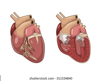
Hematopoiesis organ that produces lymphocytes. Cut-away view demonstrates structures in the heart right ventricle pulmonary trunk pulmonary arteries and lungs where adult-stage heartworms are normally found. The integumentary system is the skin and fur that cover the animals body.
Part of the digestive tract between the esophagus and the intestine. This means that for the average dog or cat everything in this diagram below happens twice each second. The axis has a dens which projects cranially to allow pivotal motion between the atlas and axis.
D49 Identify the various parts of the heart and associated major vessels. Canine In the canine heart both the right and left atrial appendages take a tubular shape and the right is generally larger than or the same size as the left 1. Leading into the right atrium are the two largest veins in the body the vena cava.
Hills Atlas of Veterinary Clinical Anatomy Normal Canine Heart Left ventricle Left atrioventricular valve Right atrioventricular valve Chorda tendinea Papillary muscle Right ventricle Right atrium Aortic arch Pulmonary artery Left atrium Coronary vessels Left ventricle Left ventricular free wall Right ventricle Right ventricular free wall Ventricular septum. There are also 2 lower chambers called the left and right ventricles. The digestive system cat dog includes the mouth teeth salivary glands esophagus stomach intestine pancreas liver and gall bladder.
From the quiz author. The dog has 321 bones. Cardiovascular and Lymphatic System.
And explain the observed latex coloration that may be present in the preserved specimen. The heart the arteries and the veins make up the closed circulatory. Canine Heart - Normal Anterior View.
There is a printable worksheet available for download here so you can take the quiz with pen and paper. The structure of the moderator band differs greatly between these hearts. Carnivorous domestic mammal raised to perform various tasks for humans.
Regions of a Long Bone Structure of a Long Bone articular cartilage nutrient artery entering nutrient foramen marrow cavity compact bone spongy bone ligament periosteum endosteum physis epiphyseal plate physis epiphyseal plate metaphysis diaphysis metaphysis epiphysis epiphysis. Seat of the intelluctual capacities of a gog. 21 Vessels of Trunk and Neck 22 The Heart 231 Cardiac Conducting System.
Unlike in the human heart the ostia of the vena cava enter the heart perpendicular to one another 8. There are upper chambers on both the left and ride sides of the heart called the left and the right atria the plural form of atrium. The spinous process is nonbifid.
Heart- The heart is responsible for pumping oxygenated blood around the body to nourish cells and promote their functions ultimately maintaining life. The heart is a hollow muscular organ which in mammals and birds is divided into 4 chambers.